What is CBCT :
CBCT or Cone Beam Computed Tomography is an advanced diagnostic imaging modality of Dental and Maxillofacial Radiology , based on computed tomography that utilizes a single rotational cone beam of xray to render 3D multiplanar images of teeth , jaw bones and adjacent vital structures of head and neck region, by reconstruction from a series of 2D projections, at reduced exposure time and lower radiation dose than medical CT .
The CBCT imaging has not only eased and improvised the diagnosis of dental & maxillofacial pathologies , but has also lead to better treatment planning for a wide range of clinical applications .
Benefits of CBCT over Medical CT :
| Conical Xray beam for volumetric data acquisition in solitary rotation |
| Limited area of Exposure by selecting a particular Field of View (FOV ) related to the area of interest |
| Improved Accuracy and Diagnostic image quality |
| Rapid scan time |
| Reduced Radiation dose |
| Decreased Image Artifacts |
| Less expensive and compact machinery |
Various indications of CBCT in dentistry are :
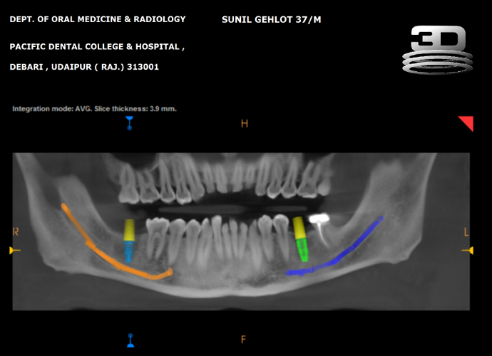
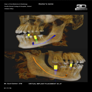
| Implant planning |
| Dental and Maxillofacial pathosis |
| Trauma cases |
| TMJ , ENT and Airway space evaluation |
| Dental / Endodontic or Periodontal Status evaluation |
| Soft tissue Calcifications of head and neck region |
| Assisting in 3D printing / Surgical guide fabrication |
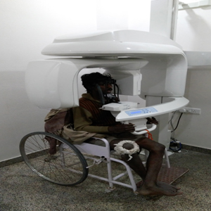
CBCT at PDCH Udaipur :
The facility of CBCT , the first to be installed in any Private Dental College in Rajasthan , is available at the Dept. of Oral Medicine and Radiology , Pacific Dental College and Hospital , Debari , Udaipur. The scans are performed on Carestream CS 9300 machine with FOV from 5X5 to 17x13.5 and voxel size from 0.09 to 0.500 mm.